


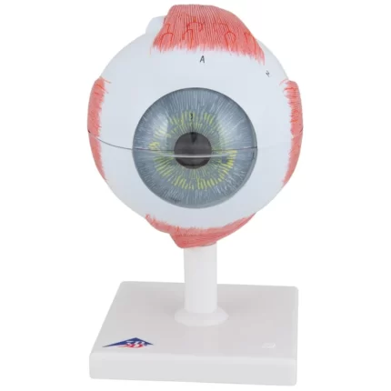

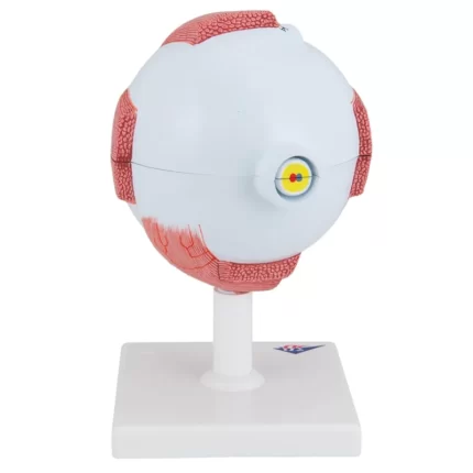
Giant Eye Model, 3 times full-size, 7 part – Includes 3B Smart Anatomy
This large anatomical human eye model shows the optic nerve in its natural position in the bony orbit of the eye (floor and medial wall).
At three times life size this eye model is great for anatomical demonstrations.
The human eyeball can be dissected into:
- Both halves of sclera with cornea and eye muscle attachments
- Both halves of the choroid with iris and retina
- Eye lens
- Vitreous humour
This high quality model is great for studying the anatomy of the human eye and the anatomy of the surrounding area! Human Eye Anatomy Model on base.


Reviews
There are no reviews yet.