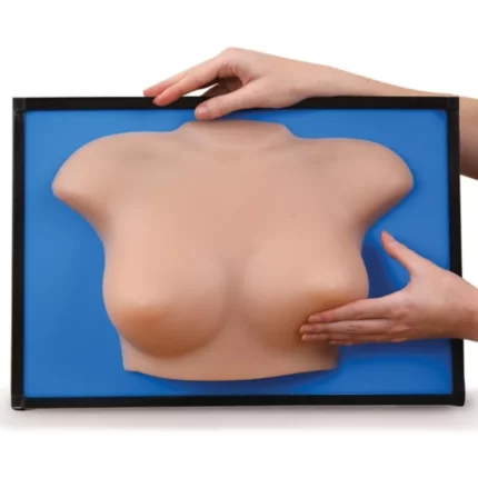


Episiotomy Suturing Simulators, Set of 3
Episiotomy Suturing Simulators, Set of 3
Now with new lifelike skin for the most realistic suturing practice!
These new and improved Life/form® Episiotomy Suturing Simulators provide a realistic way for students to learn good surgical techniques. The simulators provide students with a variety of repair experiences without the constraint of time and concern for safety, which are factors with a live patient.
The simulators can be used by the student in a learning lab with an instructor, or by the student individually in the clinical setting just prior to a patient experience. They are also useful as homework teaching aids that can be signed out at night and returned the next day. In addition to being portable, the lifelike texture allows the learner to develop a “feel” for instrument handling, tension on suture, and the advantages of one method of tying knots over another.
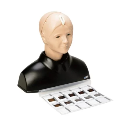
EYE Examination Simulator (3 Step)
EYE Examination Simulator (3 Step)
The EYE Examination Simulator is an innovative trainer for fundus examination, designed to allow examination of eyegrounds with the physician's own ophthalmoscope. Various cases can be set up for trainees using combinations of choice of slides, depth and pupil diameter. Soft and supple material allows hands-on simulation of real examination procedures, such as raising the eyelid.

EYE Examination Simulator II (2 Step)
The EYE Examination Simulator is an innovative trainer for fundus examination, designed to allow examination of eyegrounds with the physician's own ophthalmoscope. Various cases can be set up for trainees using combinations of choice of slides, depth and pupil diameter.
Soft and supple material allows hands-on simulation of real examination procedures, such as raising the eyelid.
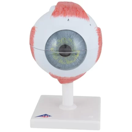
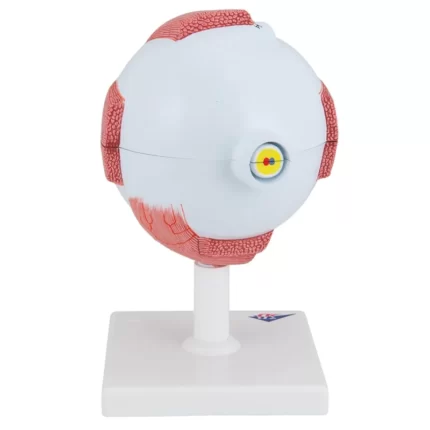
Eye Model, 5 times full-size, 6 part
Eye Model, 5 times full-size, 6 part - Includes 3B Smart Anatomy
The Giant Eye replica is a great tool to teach-learn the anatomy of the eye! Removable parts of the human eye model include:- Upper half of the sclera with cornea and eye muscle attachments
- Both halves of the choroid with iris and retina
- Lens
- Vitreous humour
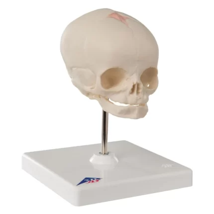
Fetal Skull Model, with stand
The anatomy of a human foetal skull can be studied easily with this replica. The skull model is a natural cast of a fetal head in the 30th week of pregnancy showing the characteristics of prenatal development. The fontanelles, which become bone over time, are clearly visible on the fetal skull. Sutures will form along the bony plates helping fuse the foetal skull as the individual ages. Foetal skull on stand.

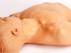
Forceps Vacuum Delivery Obstetric Manikin
Forceps Vacuum Delivery OB Manikin - Light Skin
The Forceps/Vacuum Delivery OB Manikin is used in "Advanced Life Support in Obstetrics" (ALSO) training programs. The manikin has all of the same realistic and accurate anatomical manikin features as in our Obstetrical Manikin #180, but with added features of premature and full term fetuses, Forceps and Vacuum Extractor not included. Key Features:- Soft vinyl pelvis replicates resistance encountered in a delivery requiring forceps or vacuum intervention
- Removable abdominal overlay provided is different from those that come with the #180 OB manikin, but the overlays are interchangeable, and optional
- Simulated blood powder
- Extra vulva
- Carry bag
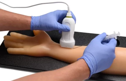
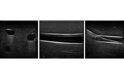
Gen II Femoral Vascular Access and Regional Anesthesia Ultrasound Training Model
- Ultrasound guided regional anesthesia and central venous access ultrasound training model
- Regional anesthesia; contains the femoral nerve and it’s branches along with the Saphenous Nerve, the Fascia Iliaca nerve
- Central venous access; contains anatomically correct vascular anatomy of the right lower torso including the femoral artery and vein, Aorta, IVC, Common Iliac Artery and Vein, Internal Iliac Artery and Vein, Great Saphenous Vein, Simulated DVT (Deep Vein Thrombosis) in the lower Left Femoral Vein.
- Inject simulated anesthetics to verify needle tip location and to practice the entire regional anesthesia procedure
- Anatomical landmarks include: Pubic Symphysis, Iliac Crest and Inguinal Ligament for palpation.
- Use 18-21 gauge needles and 7 French Catheter to access the venous system.
- Use repeatedly; injected simulated anesthetics are automatically expelled
- Superb ultrasound imaging characteristics; learn to acquire and interpret ultrasonographic imaging of nerves and vessels
- New Blue Phantom Integrated Hand pump to create pulsatile arteries with slow return to allow collapsing of the venous system
- Self healing tissue will withstand tremendous use and will save you money by dramatically reducing the necessity for purchasing replacement parts
- Utilize traditional anatomical landmarks for blind insertion techniques, or utilize ultrasound to obtain images of anatomical structures
- Accommodates full threading of guidewires and catheters
- Venous and arterial fluids that are removed during central catheter insertions training are easily refilled using quick fill ports
- Positive fluid flow in the vessels provides users with immediate feedback when vessels are accessed
- Simulated blood fluids in the arterial vessels differ from the venous system allowing for users to easily verify successful venous access procedures
- Tissues match the acoustic characteristics of real human tissue so when you use your ultrasound system on our training models, you experience the same quality you expect from imaging patients in a clinical environment
- Performs well using any ultrasound imaging system
- Practice using ultrasound system controls
- Purchase includes 2 bottles of simulated blood refill solution; one red (arterial), one blue (venous). 235mls bottles
- Model comes with Thermoform Platform to keep model in place and for storage purposes.
- High quality
- No special storage needs
- Patented technology
- Made in USA
Gen II PICC with IV & Arterial Line Vascular Access Ultrasound and X-Ray Trainer
Blue Phantom’s Second Generation PICC with I.V. and Arterial training model is designed for users interested in developing and practicing the skills associated with ultrasound guided PICC line placement, arterial line placement, and peripheral IV access. This Arterial, I.V. and PICC training model is intended to assist users in learning to place needles, guidewires and catheters in the brachial vein, basilic vein, radial artery, ulnar artery and superficial veins.
Gen II Ultrasound Central Line Training Model
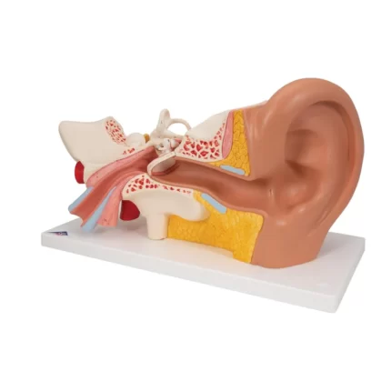
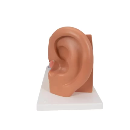
Giant Ear Model, 3x Life Size, 4-part
Giant Ear Model, 3x Life Size, 4-part - Includes 3B Smart Anatomy
This high quality model of the human ear represents outer, middle and inner ear. The detailed human ear model has removable eardrum with hammer, anvil and stirrup as well as 2-part labyrinth with cochlea and auditory/balance nerve. Ear on base for easy display in a classroom or doctor's office. This ear model is a great way to teach and study the anatomy of the human ear!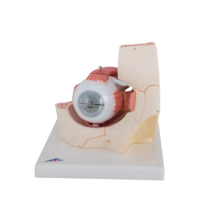
Giant Eye Model, 3 times full-size, 7 part
Giant Eye Model, 3 times full-size, 7 part - Includes 3B Smart Anatomy
This large anatomical human eye model shows the optic nerve in its natural position in the bony orbit of the eye (floor and medial wall). At three times life size this eye model is great for anatomical demonstrations.The human eyeball can be dissected into:
- Both halves of sclera with cornea and eye muscle attachments
- Both halves of the choroid with iris and retina
- Eye lens
- Vitreous humour